A medical endoscope is a tube with a light source. It can enter the human body through the natural cavity of the human body or a small incision in minimally invasive surgery, thereby helping doctors diagnose diseases or assist in surgical operations. At present, the medical endoscope system is mainly composed of three modules: the endoscope body, the image processing center, and the monitor. It is a precision detection instrument that integrates multidisciplinary knowledge such as traditional optics, modern electronics, software algorithms, ergonomics, and precision machinery. In the era of minimally invasive diagnosis and treatment, endoscopes are one of the most important development directions. Whether from the perspective of technological development, clinical needs, or policy guidance, they deserve the market's continued attention.
Image sensor
As one of the key components of the camera system, the image sensor can be divided into two types: photoconductive camera tubes and solid-state image sensors. Solid-state image sensors are further divided into charge-coupled device image sensors (CCD) and complementary metal oxide semiconductor image sensors (CMOS). Both image sensors realize their functions by converting optical signals into electrical signals.
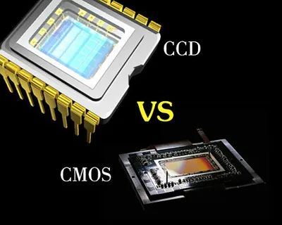
In electronic endoscopes, the image sensor is located at the front end of the camera system; in rigid tube endoscopes and optical fiber endoscopes, the image sensor is located at the rear handle. The main comparison between them is as follows:

The two have their own advantages and disadvantages in terms of performance characteristics, cost, etc., and together provide support for endoscope image acquisition.
Optical imaging system
The optical imaging system is a common part of the three major types of endoscopes and an indispensable component of fluorescent electronic endoscopes. It consists of a set of lenses, and the lens material is usually glass or artificial resin. The optical imaging system undertakes the important task of presenting the image to be photographed on CMOS or CCD.
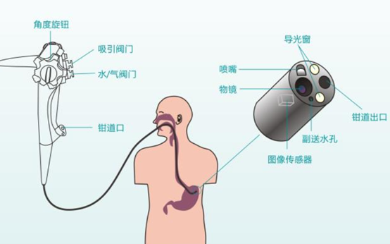
Electronic mirror structure image source network
The main parameters of the optical imaging system include field of view angle, field of view, aperture, resolution and depth of field.
Field of view angle
It refers to the angle between the incident light of the object image and the horizontal line after it first passes through the objective glass and enters the objective lens, that is, the angle of view (DOV, Direction Of View). The endoscope viewing angle is generally 0°, 12°, 30°, 70°, 90°, etc.
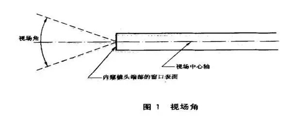
The front ends of medical electronic endoscopes and optical fiber endoscopes can be bent, but optical fiber endoscopes are far less tough than metal cables because the optical fiber is easy to break. The maximum bending angle of medical electronic endoscopes is 270°. When the field of view of the optical system is 90°, it can ensure that there is no blind spot in observation.
Field of View (FOV)
Refers to the range that the objective lens can observe, which is called the field of view. The field of view of the endoscope is generally divided into wide angle and standard angle. Different fields of view are suitable for different inspection scenarios, providing doctors with a more comprehensive observation perspective.
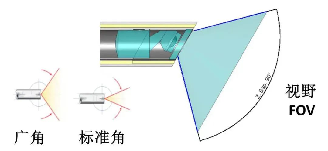
Photo source: Xueli, official account
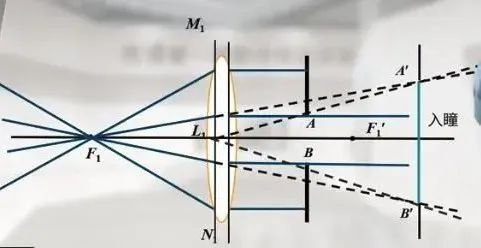
Clear Aperture
The size of the clear aperture has an important influence on the depth of field and lighting ability of the optical imaging system. The larger the clear aperture, the stronger the lighting ability of the optical imaging system, the brighter and clearer the image, and the lower the required light source illumination brightness. However, as the clear aperture increases, the depth of field of the system will decrease.
Optical imaging system resolution
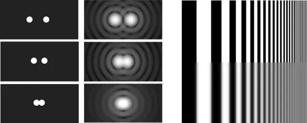
The resolution of an optical imaging system refers to the minimum distance at which the "image" of an "object" can be distinguished in detail after passing through the optical system. Two image points greater than this distance can be identified as two points, while two points less than this distance will be identified as one point after passing through the optical system. The optical resolution of an image sensor is expressed by the number of black and white line pairs that can be distinguished per millimeter, that is, there are multiple pairs of pixels per millimeter of width. It should be noted that the image resolution we conventionally understand is the number of pixels used to display the image within a unit distance.

Resolution board type A
For example, the pixels of the micro-image sensor used in smartphone cameras are as high as 20 million or even 40 million, but from the perspective of shooting effects, photography enthusiasts still often shoot with a SLR instead of a mobile phone. The main reason is that the resolution of the lens module of the optical imaging system matched by the mobile phone camera cannot reach the optical resolution of the corresponding image sensor. Therefore, the key to determining the resolution of the entire camera shooting lies in the optical imaging system.
Depth of field
That is, the clear depth of the image of the scene, which is a distance from near to far that the optical system can clearly observe. Within this distance, objects in the scene can form a clear image no matter they are moved closer to the lens or farther away. Reasonable depth of field settings can allow doctors to better observe lesions at different distances during the examination process and improve the accuracy of diagnosis.

Contact Us:
Website:www.smartveingroups.net
Tel:+86 17357337776
Email:chu@smartvein.com.cn
https://mp.weixin.qq.com/s/NPpwfqusP-YuD3TQ0iacyw
www.smartveingroups.net
SmartVein Medical Technology